Course
Kaposi’s Sarcoma
Course Highlights
- In this Kaposi’s Sarcoma
course, we will learn about the incidence rate and pathophysiology of Kaposi sarcoma. - You’ll also learn the risk factors and most common signs and symptoms of Kaposi sarcoma.
- You’ll leave this course with a broader understanding of knowledge of the complications and prognosis of Kaposi sarcoma to nursing interventions to care for clients diagnosed with the condition.
About
Contact Hours Awarded: 2
Course By:
Joanna Grayson, BSN, RN
Begin Now
Read Course | Complete Survey | Claim Credit
➀ Read and Learn
The following course content
Introduction
Kaposi sarcoma is a soft-tissue tumor caused by an infectious agent that occurs in clients with immunosuppression, particularly those with acquired immunodeficiency syndrome (AIDS) or those undergoing organ transplantation [2, 4]. The disease was first described by a Hungarian dermatologist, Moritz Kaposi, of the University of Vienna, in 1872 when he assessed pigmented cutaneous sarcoma in the lower extremities of a client [5, 10].
Kaposi sarcoma is an AIDS-defining cancer, as are liver, anal, lung and Hodgkin lymphoma [2, 4]. Male homosexuals who are HIV positive have a fivefold to tenfold increased risk of Kaposi sarcoma, and in HIV clients who are not taking high-activity antiretroviral therapy (HAART), up to 30% will develop Kaposi sarcoma [4, 5].
Over five percent of clients who undergo an organ transplant, and who subsequently develop cancer for the first time, will develop Kaposi sarcoma at an increased risk of 500-fold over the general population [4, 5]. Clients who undergo solid organ transplantation have a higher risk of developing Kaposi sarcoma than their counterparts who have bone marrow or peripheral blood stem cell transplants [4].
In the 1980s and 1990s, Kaposi sarcoma was punctuated by a five-year survival rate of roughly 10% and thus was associated with considerable morbidity and mortality [5]. Today, the survival rate ranges from approximately 70% to 95% [5]. The international incidence rate of soft tissue sarcoma ranges from roughly two to five cases per 100,000 individuals per year, with the annual rate of new cases in the United States being 3.4 per 100,000 people [9]. However, Kaposi sarcoma is a major public health concern in Eastern and Central Africa, and the challenge is compounded by the lower socioeconomic status of the region [14].
Kaposi sarcoma is a chronic condition marked by periods of relapse that require sustained systemic treatments that can lead to increased risk of toxicities, poor tolerability, and decreased quality of life [5]. Even though the disease is not common, it can cause disfigurement of the skin and death in clients, thus necessitating nursing knowledge of the condition.

Self Quiz
Ask yourself...
- In which client populations is Kaposi sarcoma most prevalent?
- What is the difference in incidence rates of clients who are diagnosed with Kaposi sarcoma and taking high-activity antiretroviral therapy versus those clients who are not taking the therapy?
- Can you compare the survival rates of Kaposi sarcoma in the 1980s and 1990s to those of today?
- How do the international rates of Kaposi sarcoma differ from those rates of the condition in the United States?
Pathophysiology
Kaposi sarcoma is a condition of malignant tumors that can arise in any organ system; it is caused by immunosuppression that permits human herpesvirus-8 (HHV-8) to interfere with normal cell function [4,5,9]. However, the specific pathophysiology is unknown since not all clients who present with HHV-8 develop the condition [5]. HHV-8 is a large, double-stranded DNA virus that belongs to the herpesvirus family, and it expresses a protein that is necessary for its replication called latency-associated nuclear antigen (LANA).
HHV-8 is transmitted via saliva, semen, intravenous drug use, and blood transfusion (Batash, Bishop). It infects cells, causing immune suppression and inflammation that permit the virus to proliferate [4]. Interestingly, the dermal lymphatic alterations associated with impaired immunity that predispose clients to HHV-8 infection can be due to exposure to volcanic soil, iron, aluminosilicate, clay, and some bloodsucking insects [5].
Conditions associated with Kaposi sarcoma are plasmablastic multicentric Castleman disease, primary effusion lymphoma, intravascular large B cell lymphoma, angiosarcoma, and inflammatory myofibroblastic tumor [4].
Kaposi sarcoma is delineated into four subtypes. These subtypes include [3,5,8,10,14]:
- Classic—This type was originally described by Kaposi and presents in middle or old age, particularly in immunocompetent men of Mediterranean and Eastern European descent.
- Endemic—This includes several forms found in individuals from sub-Saharan Africa before the AIDS epidemic. It also occurs in children in Africa, particularly in ages 4 to 9. Endemic Kaposi sarcoma is typically mild in adults, but in children, it is aggressive and can quickly metastasize to the visceral organs.
- Iatrogenic—This type is associated with immunosuppressive medication therapy, particularly in clients who have undergone organ transplantation, and those who have gastrointestinal inflammatory disease and rheumatologic conditions. Clients who take systemic corticosteroids and chemotherapy agents are also at risk.
- HIV/AIDS association—This type is linked mainly to homosexual men who are diagnosed with HIV or AIDS.
A more recent subtype of Kaposi sarcoma has developed due to increased cases of men who have sex with men (MSM) without HIV infection [5,8]. This type differs from classic Kaposi sarcoma because it develops at a younger age and has a milder presentation that does not require systemic treatment [5].
Self Quiz
Ask yourself...
- What is the pathophysiological process of Kaposi sarcoma?
- How is HHV-8 transmitted?
- Which conditions are associated with Kaposi sarcoma?
- What are the four subtypes of Kaposi sarcoma?
Risk Factors
Kaposi sarcoma occurs mainly in male clients of Eastern European and Mediterranean descent who are over 50 years of age [3,8,9]. The median age at diagnosis is 70 years, and men of Italian, Greek, and Jewish heritage are affected more than clients of other cultures [5]. The incidence of the condition has a male-to-female ratio of 17:1 [4].
Soft tissue sarcomas like Kaposi sarcomas have several risk factors. These include [3,9]:
- Contact with salicaceous volcanic soil
- Bloodsucking insect bites
- Chronic lymphedema
- Previous radiation therapy
- Arsenic, thorium dioxide, and vinyl chloride exposure
- HHV-8 infection
- HIV
- Li-Fraumeni syndrome
- von Recklinghausen disease
- Gardner syndrome
- Nevoid basal cell carcinoma syndrome
- Tuberous sclerosis
- Werner syndrome
Self Quiz
Ask yourself...
- What is the cultural profile of most clients who are diagnosed with Kaposi sarcoma?
- Exposure to which substances can increase the risk of Kaposi sarcoma?
- Which syndromes are risk factors for Kaposi sarcoma?
- Which type of medical treatment procedure can be a risk factor for Kaposi sarcoma?
Signs and Symptoms
Kaposi sarcoma is a vascular lesion that forms pinkish purple to reddish blue to brownish-black macules, plaques, and nodules on the skin’s surface or mucocutaneous surfaces [3,8,10]. These lesions can range in size from very small to several centimeters in diameter; they can grow slowly over months to years, or quickly within weeks, in some cases [8].
The three main skin presentations of Kaposi sarcoma are [4,12]:
- Patch: Flat, reddish-violaceous, well-defined margins, scaly surface
- Plaque: Violaceous-brown in color with underlying vascularity, well-defined margins, whitish scales
- Nodule: Deep purple with underlying active vascularity, rainbow pattern of colors, whitish scales
Various terms used to describe Kaposi sarcoma growths include patch, plaque, nodule, lymphadenopathic, exophytic, infiltrative, ecchymotic, telangiectatic, keloidal, cavernous, and lymphangioma-like [8].
Anatomical distribution of the lesions in adults includes the extremities (45%), intra-abdominal organs (38%), trunk (10%), and head and neck (5%) [9]. The lesions can be painful and cause lymphedema and secondary infection. They can also lead to hemorrhage. Additionally, the lesions can ulcerate and invade nearby tissues, including the lymph nodes, lungs, gastrointestinal system, and other visceral organs [4,5]. When the lungs are infected, respiratory compromise can lead to death [4].
When the lesions invade the mucous membranes of the mouth and throat, swelling in the hard palate, protruding lips, tumors in the oral cavity, and swallowing difficulties can be present [6]. Oral lesions can also grow on the gums and cause tooth displacement.
Since Kaposi sarcoma is considered a rare condition, the lesions can be misdiagnosed as arterial insufficiency or venous stasis [3]. Kaposi sarcoma can also be confused as hemangioma, venous lake, glomus tumor, pyogenic granuloma, purpura, nodular melanoma, angiosarcoma, Merkel cell carcinoma, or bacillary angiomatosis [5,12].
Signs and symptoms that accompany the dermatologic presentation of lesions in Kaposi sarcoma are unexplained fever, weight loss, and lymphadenopathy [5].

Self Quiz
Ask yourself...
- Which integumentary colors and dermatological presentations are associated with Kaposi sarcoma?
- Which terms are used to describe Kaposi sarcoma growths?
- What is the anatomical distribution of Kaposi sarcoma?
- Which organ systems can be affected by Kaposi sarcoma lesions that ulcerate?
Nursing Assessment
The nurse should take the client’s health history and perform a body systems assessment that emphasizes the skin and mucocutaneous surfaces in clients suspected of or diagnosed with Kaposi sarcoma. Purplish lesions and enlarged lymph nodes should be biopsied and sent to pathology for evaluation [4]. The nurse should also assess the client for fever and weight loss, as well as lesions in the oral mucosa.
Standard classifications like Tumor, Node, and Metastasis (TNM) are not beneficial for clients with Kaposi sarcoma since the condition typically progresses slowly and most clients present with the skin disease without other symptoms [5]. However, clients with AIDS-related Kaposi sarcoma are staged according to the AIDS Clinical Trials Group (ACTG) system.
The ACTG staging system is as follows [1,5]:
Tumor (T)
- T0: Limited disease involving localized regions of the skin, lymph nodes, or oral mucosa
- T1: The presence of edema and ulceration that indicates extensive mucosal and visceral disease
Immune System (I)
- I0: CD4 cell count greater than, or equal to, 200/µL (normal range, 600–1500/µL)
- I1: CD4 cell count less than 200/µL
Systemic Illness (SI)
- S0: No systemic illness or history of opportunistic infection or thrush
- S1: Systemic illness or history of opportunistic infection or thrush
There are some non-standardized proposed staging systems for Kaposi sarcoma, one of which is as follows [5]:
- Stage I: Greater than 15 cutaneous lesions, or involvement restricted to one bilateral anatomic site and few gut nodules
- Stage II: Both exophytic destructive lesions and locally infiltrative cutaneous lesions present
- Stage III: Widespread lymph node involvement with or without skin lesions, but no visceral organ involvement
- Stage IV: Widespread disease with multiple visceral organ involvement
Another non-standardized staging system includes [1,3,10]:
- Stage 1: Maculonodules on the lower extremities
- Stage 2: Plaques and small nodules on the lower extremities
- Stage 3: Ulcerated multiple angiomatous plaques and nodules on the lower extremities
- Stage 4: Multiple plaques and angiomatous nodules extending beyond the lower extremities
Stages 1 and 2 typically have a slow clinical course, while stages 3 and 4 can be aggressive. Stages 3 and 4 involve the viscera and gastrointestinal tract, with the lesions in stage 3 remaining on the limbs, but in stage 4 they progress to the trunk and head. Organ damage can occur during stage 3 when the viscera become involved [1].
Pediatric-specific staging of Kaposi sarcoma, called Lilongwe, includes [1]:
- Stage 1: Mild Kaposi sarcoma with lesions on the skin and mouth
- Stage 2: Lymphadenopathic Kaposi sarcoma that involves the lymph nodes with facial edema, oral nodular lesions, and lesions across the body
- Stage 3: Called woody edema Kaposi sarcoma, this stage divides clients into two groups: edema in less than ten percent of the body and edema in greater than ten percent of the body.
- Stage 4: Disseminated skin and/or visceral Kaposi sarcoma is present.
Nurses can use digital photography during the initial assessment to establish a baseline and during treatment to track the effectiveness of the therapeutic modalities [5].
Self Quiz
Ask yourself...
- For which signs and symptoms, in addition to skin lesions and enlarged lymph nodes, should the nurse assess the client?
- When ulcerated multiple angiomatous plaques and nodules are found on a client’s lower extremities, which organ system is also typically involved?
- To which body regions beyond the lower extremities do multiple plaques and angiomatous nodules extend?
- Woody edema Kaposi sarcoma is identified in which stage and in which client population?
Diagnostics and Treatment
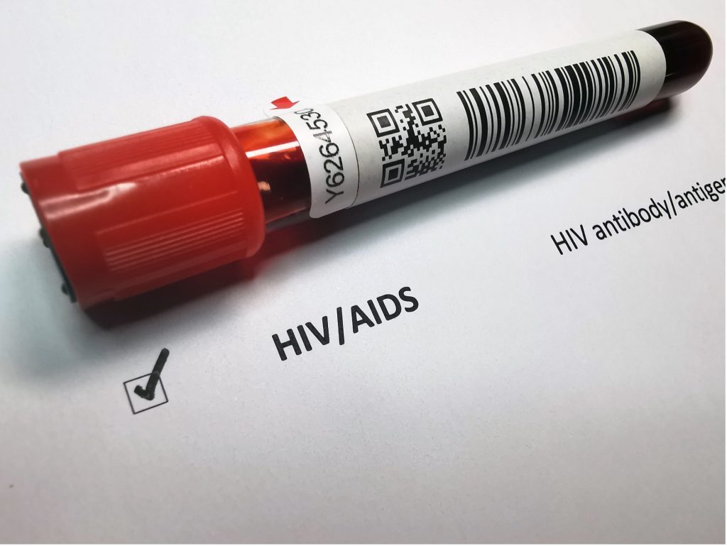
Diagnosis and treatment of Kaposi sarcoma are considered difficult by many healthcare professionals because there is not a standard set of therapeutic recommendations to support the rarity of the disease since it mainly affects a smaller population, namely HIV-positive clients [5]. All clients suspected of Kaposi sarcoma should be tested for HIV infection, and those with confirmed HIV should undergo CD4 lymphocyte count and plasma HIV viral load testing [5].
Additional tests include complete blood count (CBC), serum protein electrophoresis, C-reactive protein (CRP), erythrocyte sedimentation rate (ESR), and fecal occult blood testing (preferably three samples) to rule out concomitant gastrointestinal involvement. HHV-8 serology helps confirm the diagnosis, but not many laboratories perform this test [5]. However, plasma HHV-8 load testing can be a cost-effective measure for determining which clients should undergo more costly diagnostics, such as computed tomography (CT), positron emission tomography (PET), and biopsy. This testing can also help exclude other related disorders like Castleman’s disease and primary effusion lymphoma.
Dermatoscopy can reveal lesions with a scaly surface and rainbow pattern where several colors of the rainbow are juxtaposed. Doppler ultrasonography reveals increased vascularity within the well-defined lesions [5,12].
Core-needle or incisional biopsy is the main diagnostic mode for Kaposi sarcoma. Incisional biopsy is reserved for clients whose previous core-needle biopsies were nondiagnostic or when anatomical constraints prevent the safe performance of a core-needle biopsy [9]. The nurse should plan the initial biopsy with the surgeon, radiation oncologist, and interventional radiologist. The extracted tissues should be evaluated by a pathologist with experience diagnosing sarcomas.
Imaging is conducted before the biopsy to determine metastases. Plain radiography, CT, magnetic resonance imaging (MRI), PET, and bone scans are used. In recurrent sarcoma, CT and PET imaging have higher sensitivity than contrast-enhanced CT imaging [9]. A whole-body CT scan is used in aggressive cases to determine lymph node, viscera, bone, and liver involvement, which is common. MRI helps delineate soft tissue and bone involvement while PET scans help with whole-body staging and disease response to treatment [5].
Clients diagnosed with Kaposi sarcoma should undergo esophagogastroduodenoscopy and otorhinolaryngological evaluation, including flexible endoscopy, chest X-ray, and complete abdominal ultrasound to determine extracutaneous involvement. Endoscopy is the gold standard for diagnosing gastrointestinal lesions since they are often difficult to detect due to their small size and submucosal presentation [5]. Clients with a positive fecal occult blood test should have a colonoscopy performed, and those with clinical lymphadenopathy should undergo ultrasound testing.
The stage and severity of Kaposi sarcoma determines the treatment method. Treatment also takes into consideration the client’s medical history, physical and psychological characteristics, and comorbidities. The interdisciplinary team that guides treatment should include dermatologists, oncologists, radiation oncologists, infectious disease specialists, mental health professionals, and nurses. Surgery is not the typical treatment for Kaposi sarcoma since it is a systemic disease with multi-site involvement; therefore, surgery is relegated to palliative therapy or cosmetic purposes [3].
HAART can delay or prevent HIV from progressing to AIDS and is largely responsible for the drop in the incidence of Kaposi sarcoma [2]. In some cases, clients are treated with a combination of HAART and chemotherapy. Radiation is used to treat early stages and can be used palliatively in late stages to reduce bleeding, edema, and pain. Chemotherapy includes bleomycin, doxorubicin, etoposide, gemcitabine, paclitaxel, vinblastine, vincristine, and vinorelbine treatments. Additionally, chemotherapy is the preferred treatment for children [3,4,5,7,10]. Paclitaxel is effective in treating non-HIV/AIDS-related Kaposi sarcoma as both a first- and second-line treatment option [10,11]. Side effects of chemotherapy include alopecia, anemia, arthralgia, myalgia, chronic fatigue syndrome, onycholysis, nausea and vomiting, leukopenia, neutropenia, hand-feet syndrome, and peripheral neuropathy.
Vincristine can be injected into a large number of lesions in the same session, making it a cost-friendly treatment that is 95% effective after a single injection [5]. Bleomycin can also be used in this manner, but it is associated with a higher incidence of pain at the injection site compared to vincristine. Interferon alpha-2 can be injected into lesions twice a week and has been shown to reduce lesion size, thickness, and consistency [5].
Antiangiogenic agents pazopanib, thalidomide, pomalidomide, lenalidomide, bevacizumab, and imatinib interfere with the network of blood vessels that tumors rely on to grow and metastasize [3]. Immunotherapy agents nivolumab and ipilimumab are effective in treating Kaposi sarcoma but can carry severe complications, such as colitis, pneumonia, and lipase elevation.
Neodymium: YAG (Nd: YAG) laser treatments have an effectiveness rate of 80% in HIV-positive clients with cutaneous localized lesions [5]. Lesion size has been reduced, and elevated lesions flattened due to the Nd: YAG laser [5].
Topical therapy is used in clients with mild, localized Kaposi sarcoma and who are not good candidates for more aggressive treatments. Alitretinoin, imiquimod, and timolol creams are used, and alitretinoin is FDA-approved for use in Kaposi sarcoma [3,4,5]. Topical treatments are especially effective in mucosal lesions or when combined with other local modalities [5]. Silver nitrate cauterization and nicotine patches are also used, but with less efficacy [5]. Cryotherapy with liquid nitrogen and carbon dioxide lasers is used to treat superficial, small lesions. Minor side effects include blistering, depigmentation, and scarring of the skin.
Several new treatment options for Kaposi sarcoma are currently being studied, such as nelfinavir, valganciclovir, selumetinib, intra-lesional nivolumab, pomalidomide with liposomal doxorubicin for HIV-related neoplasms, ipilimumab and nivolumab for advanced HIV-related neoplasms, and recombinant EphB4-HAS fusion protein [4,5,7].

Self Quiz
Ask yourself...
- Which diagnostic tests should the nurse anticipate in clients with confirmed HIV diagnosis who are suspected of Kaposi sarcoma?
- Which physical characteristics of Kaposi sarcoma can dermatoscopy reveal?
- When is incisional biopsy an appropriate measure in clients suspected of Kaposi sarcoma?
- What makes gastrointestinal lesions in Kaposi sarcoma difficult to detect?
Complications and Prognosis
The larger lesions of Kaposi sarcoma can cause painful edema and disfigurement of the skin while lung involvement can cause respiratory distress and death [4]. The side effects of chemotherapy include cardiac toxicity, neuropathy, neurotoxicity, and infertility, while radiation therapy causes skin dryness, avascularity, impaired wound healing, lymphedema, and additional malignancies.
Poor prognosis is associated with age older than 60 years, tumors larger than five centimeters, high histological grade of the tumor, advanced pathological stage of the tumor, and positive tumor margins after surgery. Surgery alone is typically sufficient to cure small, low-grade tumors of the extremities or trunk while higher-grade sarcomas with higher local treatment failure rates are associated with increased metastatic potential [9]. Death occurs in 10% to 20% of clients diagnosed with Kaposi sarcoma, but a higher percentage develops fatal secondary malignancy [4]. CD4 counts and opportunistic infections are the most significant contributing factors to prognosis in Kaposi sarcoma, with lung involvement having the lowest positive outcomes [4].
Self Quiz
Ask yourself...
- Which side effects of chemotherapy and radiation in Kaposi sarcoma are most concerning?
- Which factors lead to a poor prognosis in Kaposi sarcoma?
- Which factors are considered the most significant concerning prognosis in Kaposi sarcoma?
- Which clients diagnosed with Kaposi sarcoma have the poorest outcomes?
Nursing Interventions
Since Kaposi sarcoma is mainly associated with HIV/AIDS in the United States, the nurse needs to provide client education about the prevention and control of retroviral infection. Suppressing HHV-8 infection might also play a role in controlling Kaposi sarcoma growth, so providing education about HHV-8 is also important [7]. HIV is a preventable disease, and nurses should teach clients effective measures to reduce their risk of infection.
HIV risk can be decreased by [13]:
- Using a male or female condom during sex
- Being tested for HIV and sexually transmitted infections
- Undergoing a voluntary medical male circumcision
- Using harm reduction services for people who inject and use illicit drugs
The nurse should encourage clients with HIV/AIDS to start HAART since it strengthens the immune system. Current HAART does not cure HIV infection, but it does allow the client’s immune system to grow stronger. The nurse should remind the client that HAART must be taken every day for the remainder of the client’s life.
Nurses can also implement the following interventions:
- Listen to the client’s concerns and fears and utilize therapeutic communication.
- Assist the client in a more comfortable position if the lesions are causing pain.
- Administer pain medication, if indicated.
- Provide high-calorie, high-protein meals to assist with wound healing.
- Apply compression stockings to control edema of the lower extremities. Stockings can also decrease the severity of the lesions.
- Provide rest periods and distraction techniques to promote relaxation.
- Inspect the client’s skin and mucous membranes every shift. Document new lesions.
- Monitor for side effects of chemotherapy and radiation.
- Monitor gastrointestinal and respiratory function.
- Offer emotional support to the client and their loved ones.

Self Quiz
Ask yourself...
- Why is it important for nurses to provide education about HIV/AIDS to clients diagnosed with Kaposi sarcoma?
- Which measures can clients take to reduce their risk of HIV/AIDS?
- Which important information about HAART should the nurse remind the client?
- Which type of diet can assist in healing the wounds associated with Kaposi sarcoma?
- Which cancers are considered AIDS-defining?
- In which region of the world is Kaposi sarcoma a major public health concern?
- Why is the exact pathophysiology of Kaposi sarcoma unknown?
- Which environmental factors affect the dermal lymphatic alterations associated with impaired immunity that predispose clients to HHV-8 infection?
- How do the men who have sex with men without HIV infection type of Kaposi sarcoma differ from classic Kaposi sarcoma?
- What is the male-to-female ratio of clients diagnosed with Kaposi sarcoma?
- With which conditions can Kaposi sarcoma be confused?
- Which signs and symptoms accompany the dermatologic presentation of Kaposi sarcoma?
- Why are standard classification systems not beneficial in clients with Kaposi sarcoma?
- Which classification system is used in clients with AIDS-related Kaposi sarcoma?
- A CD4 cell count of less than 200/µL corresponds with which stage of the AIDS Clinical Trials Group (ACTG) system?
- Why is digital photography a useful assessment tool to nurses caring for clients with Kaposi sarcoma?
- Clients suspected of Kaposi sarcoma should be tested for which potential underlying condition?
- Which interdisciplinary team members should help guide the treatment in the client diagnosed with Kaposi sarcoma?
- When is surgical intervention implemented in Kaposi sarcoma treatment?
- Which medication is used as both a first- and second-line chemotherapy treatment option in clients diagnosed with Kaposi sarcoma?
- Which side effects of chemotherapy should the nurse assess in clients diagnosed with Kaposi sarcoma?
Conclusion
Kaposi sarcoma is a rare type of skin cancer that is mainly associated with HIV/AIDS. The skin, mucous membranes, and major organs, such as the liver and lungs can be involved. Even though the condition has declined significantly since the 1980s and 1990s, nurses need to recognize the lesions during assessment and implement effective interventions since the condition can be fatal in some cases.
References + Disclaimer
- Addula, D., Das, C.J., Kundra, V. (2021). Imaging of Kaposi sarcoma. Abdominal Radiology, 46, 5297–5306. doi:10.1007/s00261-021-03205-6
- American Cancer Society (ACS). (2024). Cancer facts and figures 2024. Retrieved from https://www.cancer.org/content/dam/cancer-org/research/cancer-facts-and-statistics/annual-cancer-facts-and-figures/2024/2024-cancer-facts-and-figures-acs.pdf
- Batash, R., Crimi, A., Kassem, R., Asali, M., Ostfeld, I., Biz, C., Ruggieri, P., Schaffer, M. (2024). Classic Kaposi sarcoma: Diagnostics, treatment modalities, and genetic implications – a review of the literature. Acta Oncologica, 63, 783-790. https://pmc.ncbi.nlm.nih.gov/articles/PMC11495121/
- Bishop, B.N., Lynch, D.T. (2023). Kaposi sarcoma. Retrieved from
- https://www.ncbi.nlm.nih.gov/books/NBK534839/
- Denaro, N., Indini, A., Brambilla, L., Marzano, A.V., Garrone, O., Tourlaki, A. (2024). Management and future therapeutic perspectives of classic Kaposi’s sarcoma: An evidence-based review. Retrieved from
- https://www.tandfonline.com/doi/full/10.2147/OTT.S468787#d1e242
- Farnsworth, C. (2023). What are the oral symptoms of Kaposi sarcoma? Retrieved from https://www.medicalnewstoday.com/articles/kaposi-sarcoma-oral
- Goff, C.B., Dasanu, C.A. (2023). Changing therapeutic landscape in advanced Kaposi sarcoma: Current state and future directions. Journal of Oncology Pharmacy Practice, 29(4), 917–926. https://doi.org/10.1177/10781552221148
- Krown, S.E., Singh, J.C. (2024). Classic Kaposi sarcoma: Clinical features, staging, diagnosis and treatment. Retrieved from
- https://www.uptodate.com/contents/classic-kaposi-sarcoma-clinical-features-staging-diagnosis-and-treatment
- National Cancer Institute (NCI). (2023). Kaposi sarcoma treatment (PDQ)—health professional version. Retrieved from
- https://www.cancer.gov/types/soft-tissue-sarcoma/hp/adult-soft-tissue-treatment-pdq
- Paksoy, N., Khanmammadov, N., Doğan İ., Ferhatoğlu, F., Ahmed, M.A., Karaman, S., Aydiner, A. (2023). Weekly paclitaxel treatment in the first-line therapy of classic Kaposi sarcoma: A real-life study. Medicine, 102(5), e32866. doi:10.1097/MD.0000000000032866
- Tourlaki, A., Germiniasi, F., Rossi, L.C., Veraldi, S., Brambilla, L. (2020). Paclitaxel as first- or second-line treatment for HIV-negative Kaposi’s sarcoma: A retrospective study of 58 patients. Journal of Dermatological Treatment, 31(2),183–185. https://doi.org/10.1080/09546634.2019.1590520
- Tourlaki, A., Nazzaro, G., Wei, Y., Buffon, S., Mattioli, M.A., Marzano, A.V., Brambilla, L. (2022). Clinical, dermoscopic, ultrasonographic, and histopathologic correlations in Kaposi’s sarcoma lesions and their differential diagnoses: A single-center prospective study. Journal of Clinical Medicine,12(1), 278. doi:10.3390/jcm12010278
- World Health Organization (WHO). (2024). HIV and AIDS. Retrieved from https://www.who.int/news-room/fact-sheets/detail/hiv-aids.
- Zeinaty, P.E., Lebbé, C., Delyon, J. (2023). Endemic Kaposi’s sarcoma. Cancers, 15(3), 872. doi:10.3390/cancers15030872
Disclaimer:
Use of Course Content. The courses provided by NCC are based on industry knowledge and input from professional nurses, experts, practitioners, and other individuals and institutions. The information presented in this course is intended solely for the use of healthcare professionals taking this course, for credit, from NCC. The information is designed to assist healthcare professionals, including nurses, in addressing issues associated with healthcare. The information provided in this course is general in nature and is not designed to address any specific situation. This publication in no way absolves facilities of their responsibility for the appropriate orientation of healthcare professionals. Hospitals or other organizations using this publication as a part of their own orientation processes should review the contents of this publication to ensure accuracy and compliance before using this publication. Knowledge, procedures or insight gained from the Student in the course of taking classes provided by NCC may be used at the Student’s discretion during their course of work or otherwise in a professional capacity. The Student understands and agrees that NCC shall not be held liable for any acts, errors, advice or omissions provided by the Student based on knowledge or advice acquired by NCC. The Student is solely responsible for his/her own actions, even if information and/or education was acquired from a NCC course pertaining to that action or actions. By clicking “complete” you are agreeing to these terms of use.
➁ Complete Survey
Give us your thoughts and feedback
➂ Click Complete
To receive your certificate